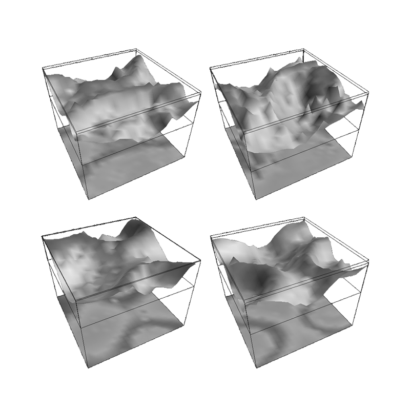
 Figure 5
Freeze-frame snapshots of surface electrocortical potential from a simulation operating at high cortical activation, under the constraint of powerful, fast, synaptic feedbacks. Frames are seperated by approximately 10 ms, and scales are normalised units. The upper, folded, surfaces show the spatial conformation of local field potentials, with increasing membrane depolarisation associated with the upward direction. The shading on this surface is present merely to enhance the visual form of the contour. The underlying flat surfaces show concurrent pulse density, and here the darker shading indicates higher pulse density, associated with greater mean membrane depolarisation. Other than an initiating impulse of spatio-temporal white noise, there is no random driving. The activity is self-sustaining.
|