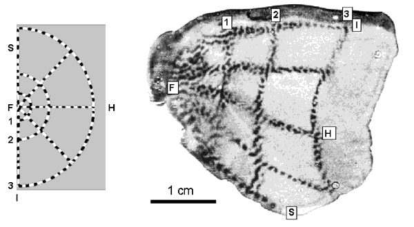
Pictorial summary of the physiology of the primary visual cortex relevant to the LGS model (see captions and sections 2 and 3 of text for details).

Global Retinotopy in Macaque: A flickering stimulus (left) and its retinotopic representation in layer 4C of V1 (right), revealed through CO staining. Reproduced from Tootell et al (1988a).
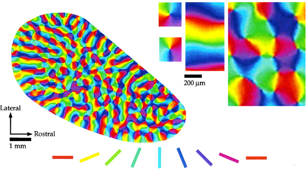
Orientation Preference in Tree Shrew: Complete map of orientation preference (left) and detail of singularities, linear zone and saddlepoints (right). Reproduced from Bosking et al (1997).
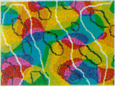
Tiling of response properties in Macaque: CO blobs (black lines), ocular dominance borders (white lines) and orientation preference (colour) tile the surface of V1 with related spatial frequencies. Reproduced from Bartfeld and Grinvald (1992).
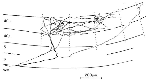
LGN Inputs to Layer 4C in Macaque: Thalamic inputs reach layer 4C in discrete blocks. Dotted lines show the borders of ocular dominance bands, with terminal boutons skipping every other band. Reproduced from Blasdel and Lund (1983).
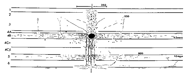
Lateral Fibres within Lamina 4B in Macaque: Staining with Horseradish Peroxidase shows long range lateral fibres extending within lamina 4B. Reproduced from Blasdel et al (1985).
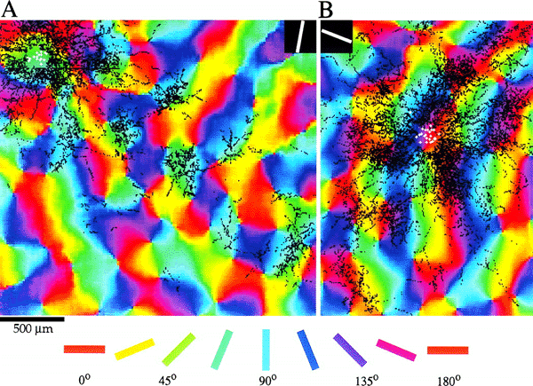
Orientation Specificity of Intrinsic Patchy Connections in Tree Shrew: Tracer injection (white dots) into a site of particular orientation preference shows axons terminating at sites of similar orientation preference (black lines). Reproduced from Bosking et al (1997).
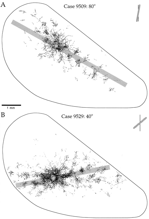
Global Pattern of Intrinsic Patchy Connections in Tree Shrew: Tracer injection into a site with particular orientation preference shows the elongation of fibre distibution in the direction corresponding to the retinotopic representation of a line of that orientation preference. Reproduced from Bosking et al (1997).